

Document Type
Dataset
Publication Date
6-2019
Data Directories
[ Wild Type, hss1a, hss1ayot, nonhsyot ] | -- Raw …..| -- AT ………| -- *.h5 ………| -- ilastik.ilp …..| -- Gfap ………| -- *.h5 ………| -- ilastik.ilp | -- Probability ………| -- AT …………..| -- *_Probabilities.h5 ………| -- Gfap …………..| -- *_Probabilities.h5 | -- PSI ………| -- AT_*.psi ………| -- Gfap_*.psi ………| -- model.csv | -- landmarks.csv
Experiment Description
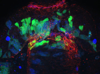
The post optic commissure (POC) is a parabolic ribbon like structure which connects the two halves of the ventral anterior nervous system. It develops in concert with a host of glial cells, called a glial bridge, which has been proposed by us and others to perform the region for POC development. We and others have shown that a set of cell guidance ligands, Slits, are required for POC development, and that disturbance of these ligands leads to a variety of POC and glial bridge defects. In particular, slit1a has been suggested to be a non-canonical guidance receptor whose influence on the POC and glial bridge remains unclear. Using the you-too (Gli2DR) slit misexpression model which exhibits reduced slit1a and increased slit2/3 and coincident loss of POC formation, we have attempted to add back slit1a to rescue POC formation. We have built ΔSCOPE as a computational analytical tool and enable quantitative analysis of rescue and loss of function experiments performed on biological structures. We have used ΔSCOPE to determine whether slit1a can rescue POC development in the you-too background. We show that slit1a alone is not sufficient to rescue POC formation, but that it has a significant effect on glial bridge formation, which may contribute to the continued POC disruption observed in the slit1a you-too addbacks.
Data Description
Raw: After collection on a Leica LSCM, data from each sample and each channel were saved individually to hdf5 files. The files are saved in a subdirectory for each channel as appropriate. Additionally, the ilastik file that was used to process the raw data is included in the subdirectory for each channel. Probability: Raw data files were processed using the ilastik training file saved in each raw channel subdirectory. The output from ilastik is a two channel hdf5 file saved to the appropriate channel subdirectory. PSI: After processing through ΔSCOPE, the resulting data is saved to a single directory as a psi file. The model file contains the coefficients that specified the quadratic function fit to each sample.
Collection Method
28 hpf zebrafish embryos from wild type (tg(hsp70:mcherry) and tg(hsp70:slit1a-mcherry) and you-too (gli2DR) homozygote mutant backgrounds (tg(hsp70:mcherry) and tg(hsp70:slit1a-mcherry) (you-too embryos were genotyped for you-too (see paper methods)) were fixed in 4% PFA for 3 hrs (or o/n 4ºC). Immunocytochemistry using mouse IgG2b anti-acetylated tubulin and mouse IgG1 anti-ZRF1 (ZIRC) (anti-Gfap) made up in PBStx (2% triton x100) with 5% BSA and 10% normal goat serum. Secondaries used were alexaflour goat anti-mouse IgG2b 488 and goat anti-mouse IgG1 594 (invitrogen). Heads were dissected and mounted for coronal collection of z-stacks imaging from most anterior of the POC to the the start of the ventral rostral cluster. Images were collected on a leica scanning confocal microscope (LSCM) SP5, using an argon 488 laser and a 633 laser. Care is taken to ensure that dichroic mirrors do not pick up signal from adjacent channels. Imaging is performed at 63x with a 1.5x digital zoom with a pixel setting of 1024 x 1024, yielding a 0.128 x 0.128 um xy dimension, and a z thickness of 0.21 um is selected. Post collection, images are imported into Fiji/Imagej, and collections are cropped to select only the immediate surrounding of the POC, approximately from the luminal junction of the telencephalon and diencephalon to a corresponding distance in the ventral direction on the other side of the POC. Care is taken to eliminate any AT signal contributions of the telencephalic anterior commissure. After cropping, any associated channels are then split, and saved separately as HDF5 files for Ilastik processing.
File Formats
HDF5: Image-based data are saved as hdf5 files, which are compatible with Ilastik. The file format is described here https://portal.hdfgroup.org/display/support. Hdf5 files can be easily viewed using the plugin for Fiji https://github.com/fiji/HDF5_Vibez/. ILP: These files are generated by the open source machine learning program ilastik https://github.com/fiji/HDF5_Vibez/. They contain the training data used to generate the probability files. PSI: Text based file format similar to a csv with space delimiters. The first twenty lines of the file specify details about the file including the columns: x, y, z, alpha, theta, r.
Recommended Citation
Barresi Lab, Smith College, "Slit1a Rescue: Role of slit1a, HSS1AYOT" (2019). Dataset, Smith College, Northampton, MA.
https://scholarworks.smith.edu/bl_ds_ros/2
Comments
This is file contains the zipped HSS1AYOT folder with Probablilty, Psi, and Raw folders within.
Additional files: .json and .csv